Home » Without Label » Anatomy Of Musckes Sndctendons - Muscle - Anatomy & Physiology Bsc2086l with Teagarden at ... : The muscles in your forearm cross the elbow and attach to the humerus.
Anatomy Of Musckes Sndctendons - Muscle - Anatomy & Physiology Bsc2086l with Teagarden at ... : The muscles in your forearm cross the elbow and attach to the humerus.
Anatomy Of Musckes Sndctendons - Muscle - Anatomy & Physiology Bsc2086l with Teagarden at ... : The muscles in your forearm cross the elbow and attach to the humerus.. All the muscles are innervated either by the medial plantar nerve or the lateral plantar nerve, which are both branches of the tibial nerve. As these muscles contract and relax, they move skeletal bones to create movement of the body. You can click the links in the image, or the links below the image to find out more information on any muscle group. The majority of muscles in the leg are considered long muscles, in that they stretch great distances. Muscles of the neck (musculi cervicales) the muscles of the neck are muscles that cover the area of the neck hese muscles are mainly responsible for the movement of the head in all directions they consist of 3 main groups of muscles:
The quadriceps and the hamstrings. When the muscle contracts, the tendons are pulled, and the bone is moved. Originates from the upper part of the fibula, passes underneath the foot and attaches by the medial foot arch peroneus brevis: It is comprised of two bones: The legs are the lower limbs of the human body that provide support and stability in addition to allowing movement.
Drawsh: November 2011 from 4.bp.blogspot.com Muscles of the neck (musculi cervicales) the muscles of the neck are muscles that cover the area of the neck hese muscles are mainly responsible for the movement of the head in all directions they consist of 3 main groups of muscles: The quadriceps and the hamstrings. A tendon connects the muscle to the bone. The muscles in your forearm cross the elbow and attach to the humerus. Lying exposed between the protective bones of the superiorly located ribs and the inferiorly located pelvic girdle, the muscles of this region play a critical role in protecting the. On the other hand, the insertion is where a tendon attaches that muscle to the *more* movable bone. The important tendons of the elbow are the biceps tendon, which is attached the biceps muscle on the front of your arm, and the triceps tendon, which attaches the triceps muscle on the back of your arm. The muscles you probably know the best are your.
All the muscles are innervated either by the medial plantar nerve or the lateral plantar nerve, which are both branches of the tibial nerve.
Take this specially designed quiz to test your knowledge about the hand and wrist. You can click the links in the image, or the links below the image to find out more information on any muscle group. Anterior, lateral and posterior groups, based on their position in the neck.the musculature of the neck is further divided into more specific groups. On the other hand, the insertion is where a tendon attaches that muscle to the *more* movable bone. The quad muscles— which form the meaty mass on the front of your thighs — are among your strongest muscle groups, and play a critical role in athletic activities. The rotator cuff is a group of four muscles and tendons that surround the glenohumeral joint. The legs are the lower limbs of the human body that provide support and stability in addition to allowing movement. Muscles of the neck (musculi cervicales) the muscles of the neck are muscles that cover the area of the neck hese muscles are mainly responsible for the movement of the head in all directions they consist of 3 main groups of muscles: The peroneal tendons run down together behind the outer side of the ankle and then split before attaching to different parts of the foot. The quadriceps muscles provide strength and power with knee extension (straightening). Originates from the upper part of the fibula, passes underneath the foot and attaches by the medial foot arch peroneus brevis: These muscles allow the ankle to bend downward and outward. Although the majority of the muscle mass is located anteriorly to the humerus, it has no attachment to the bone itself.
The peroneal tendons run down together behind the outer side of the ankle and then split before attaching to different parts of the foot. The rotator cuff is a group of four muscles and tendons that surround the glenohumeral joint. The peroneal muscles (peroneus longus and peroneus brevis), on the outside edge of the ankle and foot. Originates from the lower part of the fibula and attaches to the outer side of the midfoot You can click the links in the image, or the links below the image to find out more information on any muscle group.
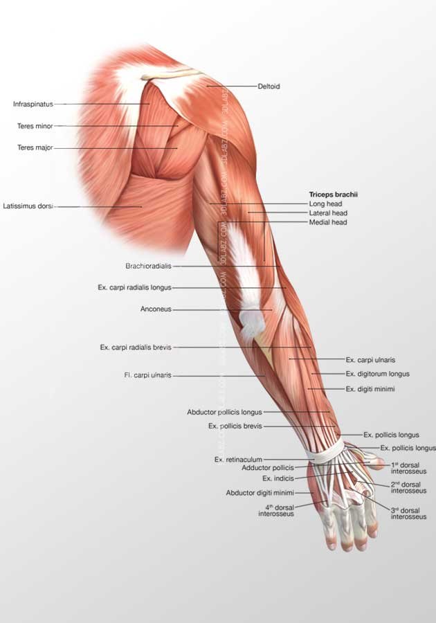
Arm Posterior Muscles 3D Illustration from www.3dlabz.com The quadriceps are a collection of 4 muscles on the front of the thigh and are responsible for straightening the knee by bringing a bent knee to a straightened position. The peroneal tendons run down together behind the outer side of the ankle and then split before attaching to different parts of the foot. Take this specially designed quiz to test your knowledge about the hand and wrist. The calf muscles (gastrocnemius and soleus), which are connected to the calcaneus via the achilles tendon. The posterior upper leg muscles provide your knees with mobility (extension, flexion and rotation) and strength. Originates from the upper part of the fibula, passes underneath the foot and attaches by the medial foot arch peroneus brevis: As these muscles contract and relax, they move skeletal bones to create movement of the body. The tendons for these muscles begin at your ischial tuberosity, or ischium (the bony bump under each buttock), and attach on the outer edges of your shinbones (tibia and fibula) just below the back of your knee.
The tendons are the attachment of the muscle to the bone.
Originates from the upper part of the fibula, passes underneath the foot and attaches by the medial foot arch peroneus brevis: The legs include the upper leg, knee, lower leg, ankle, and. The hip joint is the junction where the hip joins the leg to the trunk of the body. A tendon connects the muscle to the bone. The peroneal tendons run down together behind the outer side of the ankle and then split before attaching to different parts of the foot. Major muscles of the ankle. The thigh bone or femur and the pelvis which is made up of three bones called ilium, ischium, and pubis. On the other hand, the insertion is where a tendon attaches that muscle to the *more* movable bone. Maybe you would like to learn more about one of these? The muscles in your forearm cross the elbow and attach to the humerus. Four muscles and their attached tendons make up the rotator cuff. Every skeletal muscle has three main parts: The majority of muscles in the leg are considered long muscles, in that they stretch great distances.
This video also provides you with a. Maybe you would like to learn more about one of these? Each of them aids in a specific motion of your shoulder. The tendons are the attachment of the muscle to the bone. Together, these muscles straighten your knee, stabilize your knee joint, assist in flexing your hip (drawing your knee towards your chest), and help absorb force when you land after jumping or leaping.
Muscle and Tendon Characteristics - Classic Human Anatomy ... from schoolbag.info The important tendons of the elbow are the biceps tendon, which is attached the biceps muscle on the front of your arm, and the triceps tendon, which attaches the triceps muscle on the back of your arm. This is lesson 1 on the anatomy of the forearm. The thigh bone or femur and the pelvis which is made up of three bones called ilium, ischium, and pubis. Lying exposed between the protective bones of the superiorly located ribs and the inferiorly located pelvic girdle, the muscles of this region play a critical role in protecting the. Although the majority of the muscle mass is located anteriorly to the humerus, it has no attachment to the bone itself. All the muscles are innervated either by the medial plantar nerve or the lateral plantar nerve, which are both branches of the tibial nerve. Together, these muscles straighten your knee, stabilize your knee joint, assist in flexing your hip (drawing your knee towards your chest), and help absorb force when you land after jumping or leaping. The answers to the questions are provided at the end of the book.
Muscles of the neck (musculi cervicales) the muscles of the neck are muscles that cover the area of the neck hese muscles are mainly responsible for the movement of the head in all directions they consist of 3 main groups of muscles:
Anterior, lateral and posterior groups, based on their position in the neck.the musculature of the neck is further divided into more specific groups. Muscles of the neck (musculi cervicales) the muscles of the neck are muscles that cover the area of the neck hese muscles are mainly responsible for the movement of the head in all directions they consist of 3 main groups of muscles: The tendons for these muscles begin at your ischial tuberosity, or ischium (the bony bump under each buttock), and attach on the outer edges of your shinbones (tibia and fibula) just below the back of your knee. They act collectively to stabilise the arches of the foot, and individually to control movement of the digits. Maybe you would like to learn more about one of these? The legs are the lower limbs of the human body that provide support and stability in addition to allowing movement. The answers to the questions are provided at the end of the book. The muscles of the abdomen, lower back, and pelvis are separated from those of the chest by the muscular wall of the diaphragm, the critical breathing muscle. These muscles allow the ankle to bend downward and outward. The muscles in your forearm cross the elbow and attach to the humerus. On the other hand, the insertion is where a tendon attaches that muscle to the *more* movable bone. You can click the links in the image, or the links below the image to find out more information on any muscle group. There are tendons in your elbow that attach muscle to bone.